FA
Today, Thursday، 2 Jan 2025 - 21:32
Mokhafaf

Acronym Finder

YS
What is YS ?
Yolk Sac
This term in pregnancy and fetal ultrasound means yolk sac (iodine and iodine)....
GS
What is GS ?
Gestational Sac
This term in pregnancy ultrasound means the gestational sac (GS)....
TVS
What is TVS ?
Trans Vaginal Sonogram
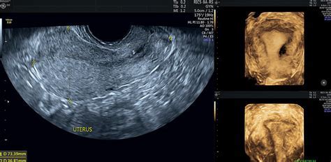
Transvaginal ultrasound iodine, is one type of ultrasound that is prescribed for women and expectant mothers. This vaginal ultrasound is very accurate in diagnosis and helps the doctor to better diagnose the disease or the patient\'s health. Transvaginal ultrasound using high-frequency sound waves, creates and shows images of the internal parts of the body. Although abdominal ultrasounds are accurate and show accurate information about the internal parts of the abdomen, they are not very accurate about the pelvic organs, and the best suggestion for examining the pelvic organs is to use a transvaginal. in transvaginal ultrasound unlike normal abdominal ultrasound where the transducer is placed on the abdomen, the respective doctor inserts an ultrasound probe, which is about 2 or 3 inches, into the vaginal canal. Doctors use this ultrasound to examine female reproductive organs such as the ovaries, erus, cervix, fallopian tubes and bladder. These organs are easily visible through the vagina.

use cases of trans-vaginal ultrasonography:
- hip pain
- bleeding, unusual vaginal
- examination, abnormal pelvis or abdomen
- a pregnancy outside the uterus
- check for cysts or fibroids uterine
- na fertility
- confirm placement IUD fit
- cancer of the reproductive organs...
ROMA
What is ROMA ?
Risk of Ovarian Malignancy Algorithm
Ovarian cancer is the fourth leading cause of cancer deaths in women worldwide and accounts for 5% of cancer deaths in women . However, there is no tool for early detection of ovarian cancer. It should be noted that 90% of breast cancers are detected using mammography in the early and curable stages of iodine, and screening of erine cancer can be done using Pap smears, which can detect early-onset cancer in the early stages. prediction of ovarian epithelial cancer (ovarian malignancy risk algorithm) can be done to women before and after and these women are classified as low-risk and high-risk groups. In women before menopause. Roma less than 11٫4 with low risk are introduced, and in women with Roma over 11٫4, and the stakes are high, or as high-risk are regarded as an. For women or women with three levels less than 9 29 9 is considered low risk, and women with R 29 9 are more likely to be identified as high-risk or high-risk ovarian cancer....
BPP
What is BPP ?
Biophysical Profile
Fetal health assessments are one of the most important concerns of mothers and fathers. Many tests are carried out in this regard regarding the possibility of various problems. One of these tests is a biophysical embryonic profile. The iodine test is one of the prenatal tests that evaluate the condition of the fetus in iodine. In this test, the fetal heart rate (by stress-free test or iodine) and ultrasound of the fetus are used to assess the heart rate ، breathing movements، muscle tone and fluid level meniotic. Then the results of iodine and ultrasound are intertwined and the score of each fetus is determined. > < < > biophysical profile is a simple and painless test performed during pregnancy to assess the health of the fetus. Especially to ensure that the fetus receives enough oxygen iodine from the erus. This iodine test is often performed when the due date of delivery has passed. Also, if you have a high-risk pregnancy this the test can also be done in the third trimester....
SIS
What is SIS ?
Saline Infusion Sonography
Ultrasonography can provide an image of the erus and possible fibroid or polyps. Saline or gel inside the erine cavity will make the ultrasound picture clearer. This method of ultrasound is called saline (iodine; iodine). Usually this picture is just two-dimensional. Nowadays, it is possible to create a three-dimensional image so that the type of anomaly can be better seen....
TUMT
What is TUMT ?
Transurethral Microwave Thermotherapy
Microwave thermotherapy through the urethra and into (uu) is one of the most effective and safe urological operations used in the treatment of benign prostatic hyperplasia (BPH). Microwave thermotherapy through the urethra provides a one-time efficient treatment for Benign Prostatic Hyperplasia. This is an alternative treatment for drugs such ASUENTAGONISTSU and prostate removal or prostatectomy. < < > microwave thermotherapy through the urethra is a non-surgical treatment that can be performed under local anesthesia on an outpatient basis. Treatment consists of inserting a special microwave urinary catheter into the urethra and in the place of the enlarged prostate. The microwave is placed inside the catheter and then the microwave emits radiation to heat and destroy the surrounding prostate tissue.< < > the procedure can take from 30 minutes to an hour and is well tolerated by patients. After the procedure-the prostate tissue will swell and irritate. The urologist often prevents the patient's urinary retention by inserting a foley catheter. After 3 to 5 days the Foley can be replaced with a temporary prostate stent, which is to improve urination without exacerbating the symptoms of inflammation.

the main risks thermography, microwave through the urethra (TUMT) include:
- retention of urine
- infection
- pain in the region of the urinary-genital.< < > evidence has shown that microwave thermotherapy operations through the urethra have been welcomed by patients more than urethral prostate scraping operations.fortunately, the recovery period after treatment is relatively quick and according to and the average recovery time is less than 5 days of rest at home. However, microwave thermotherapy through the urethra can lead to urinary retention....
IGRT
What is IGRT ?
Image Guided Radiation Therapy
IPhone a müzik (IGRT) stands for Image-guided radiation therapy is the literal meaning therapy (radiotherapy), under the guidance of the picture

the order is a method in which the computer to create an image of the tumor used to be the beam of radiation during radiotherapy directed. These images are made using ultrasound radiography (or other imaging methods. Radiation therapy can be used more precisely using the procedure and apples to healthy tissues can be reduced....